
The firm, irregular lumps that often appear weeks after surgery are not necessarily a permanent failure of your procedure, but rather a physiological crossroads between temporary fluid congestion and true scarring. It's common to feel a deep sense of anxiety when your once-smooth contours begin to feel rigid, restricted, or uncomfortably tight during the recovery process. Recognizing the early signs of fibrosis after BBL is the most critical step in preventing these adhesions from maturing into permanent, protein-rich tissue that compromises your final aesthetic results. You deserve to know whether the changes you're feeling are a standard part of the healing cycle or a complication requiring professional intervention.
This clinical guide provides the expert clarity you need to distinguish between normal post-operative edema and the development of pathological tissue adhesions. We'll examine the specific diagnostic criteria for identifying fibrosis early, the biological timeline of tissue remodeling, and the non-invasive therapeutic protocols used by specialists to restore a soft, contoured appearance. By understanding these physiological shifts, you can move forward with a clear, evidence-based plan for a successful and healthy recovery.
Key Takeaways
- Understand the biological mechanism of fibrosis, where cannula trauma triggers an overproduction of connective tissue during the inflammatory phase.
- Learn to differentiate between standard post-operative edema and the specific signs of fibrosis after BBL, including palpable "marbles" and skin pulling.
- Identify how high-volume liposuction and compression garment fitment influence the health of your lymphatic pathways during recovery.
- Discover clinical management strategies like Manual Lymphatic Drainage and the use of specialized foams to smooth tissue and reduce protein-rich fluid pockets.
- Gain insight into the necessity of a structured recovery plan guided by a clinical specialist to ensure long-term aesthetic and physiological results.
What is Post-Surgical Fibrosis? The Science of Tissue Remodeling
To understand the recovery process, we must first answer a fundamental question: What is Post-Surgical Fibrosis? In a clinical context, post-surgical fibrosis is the pathological overproduction of dense, fibrous connective tissue. This isn't a random occurrence; it's a direct response to the mechanical trauma caused by the liposuction cannula during the fat harvesting phase of a BBL. While some level of scarring is a necessary component of wound closure, fibrosis represents a healing response that's become hyper-productive and disorganized.
The primary architects of this process are cells called fibroblasts. These cells migrate to the surgical site to synthesize collagen and create a biological internal scar that stabilizes the disrupted tissue layers. However, when the inflammatory phase is prolonged, these fibroblasts produce an excessive amount of collagen that lacks the organized structure of healthy skin. This buildup creates adhesions, which are essentially internal bands of scar tissue that tether the skin to the underlying muscle or fascia. One of the primary catalysts for this transition is the presence of stagnant interstitial fluid. This protein-rich edema acts as a chemical signal that recruits more fibroblasts, effectively turning a temporary fluid issue into a permanent structural complication.
The Inflammatory Cascade and Scar Formation
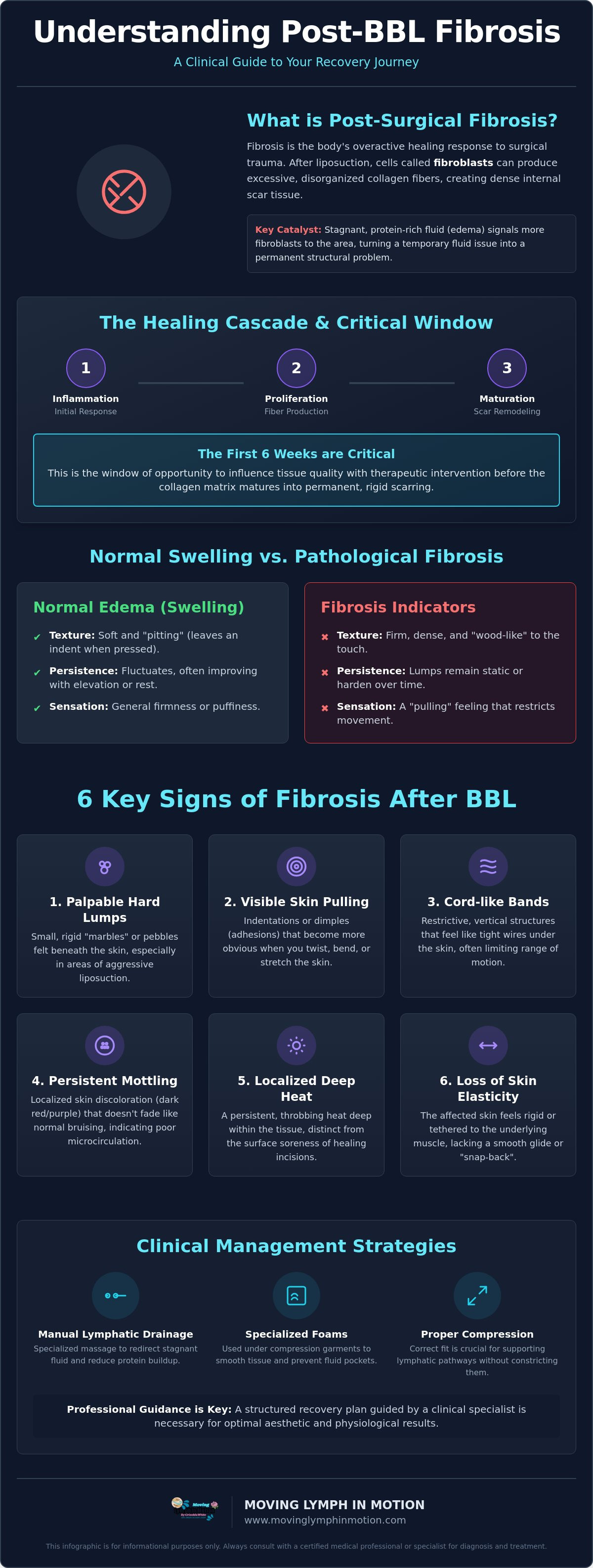
Healing follows a precise biological timeline. It starts with acute inflammation and moves into the proliferative phase within days. During this window, the body attempts to fill the void left by removed adipose tissue with dense fibers. If the lymphatic system isn't supported, the accumulation of cellular debris and proteins accelerates this fiber production. Early intervention within the first six weeks is the critical window of opportunity to influence tissue quality before the matrix matures into permanent, rigid scarring.
Normal Swelling vs. Pathological Fibrosis
Distinguishing between normal edema and the signs of fibrosis after BBL requires a careful assessment of tissue texture. While generalized firmness is expected during the first 14 days as inflammation peaks, you should monitor for these specific clinical indicators:
- Texture: Fibrosis feels firm and wood-like, whereas edema is soft and "pitting" (leaving an indentation when pressed).
- Persistence: Normal swelling fluctuates throughout the day; fibrotic lumps remain static or harden over time.
- Mobility: Adhesions cause a "pulling" sensation that restricts your natural range of motion during stretching.
Biological markers, such as increased dermal tension, indicate that the extracellular matrix is becoming overly dense. Professional assessment by a Certified Lymphatic Therapist is often necessary to disrupt this process before the tissue matures and the adhesions become more difficult to manage.
6 Key Signs of Fibrosis After BBL: What to Look For
Identifying the signs of fibrosis after BBL requires a methodical approach to self-assessment during the critical weeks of recovery. While general swelling is a systemic response to surgical trauma, fibrosis is localized and structural. It represents a shift from fluid-based congestion to organized, dense tissue. Recognizing these indicators early allows for therapeutic intervention before the collagen matrix fully matures. You should monitor your donor sites, particularly the abdomen and flanks, for these six clinical red flags:
- Palpable Hard Lumps: These often feel like "marbles" or small, rigid pebbles beneath the dermal layer. They're typically found in areas where liposuction was most aggressive.
- Visible Skin Pulling: Also known as adhesions, these appear as indentations or dimples that become more pronounced when you twist, bend, or stretch.
- Cord-like Bands: These are restrictive, vertical structures that feel like tight wires under the skin. They often limit your range of motion and prevent you from standing fully upright.
- Persistent Mottled Appearance: Unlike standard bruising that fades within 14 to 21 days, this is a localized skin discoloration that stays dark red or purple, indicating compromised microcirculation.
- Localized Deep Heat: A persistent, throbbing heat deep within the tissue that feels distinct from the surface soreness of healing incisions.
- Loss of Skin Elasticity: The affected area lacks "snap-back" and feels rigid or tethered to the underlying muscle fascia rather than gliding smoothly over it.
Effective management relies on established clinical protocols for breaking down fibrotic tissue, which are rooted in understanding how the proliferative phase of healing can veer into pathological scarring if left unsupported. If you notice these symptoms, a clinical evaluation is the next logical step to prevent permanent contour irregularities.
Tactile Indicators: How the Tissue Feels
A simple "pinch test" can help you assess tissue mobility. Gently grasp a small section of skin between your thumb and forefinger. In healthy healing tissue, the skin should lift easily away from the deeper layers. If the tissue feels "woody," leathery, or fused to the structures beneath it, fibrosis has likely begun to settle. This density often starts deep within the subcutaneous space before becoming palpable at the surface. Early detection through touch is often more reliable than visual inspection alone during the first month of recovery.
Visual and Functional Red Flags
Functional limitations are perhaps the most telling signs of advanced adhesions. If you find yourself unable to reach for overhead items or if your "faja" feels like it's digging into specific hardened ridges, the tissue is losing its natural pliability. "Shelfing," or unnatural contour lines where the skin appears to fold over a hardened internal mass, is a clear visual signal that the internal scar tissue is pulling on the surface layers. When these functional restrictions occur, consulting specialized manual lymph drainage therapists can provide the professional assessment needed to disrupt these adhesions. You may find that a clinical recovery session is necessary to restore the fluid dynamics required for a smooth, contoured result.
Why BBL Recovery Carries Unique Fibrosis Risks
The Brazilian Butt Lift (BBL) presents a dual-phase physiological challenge that differs significantly from isolated liposuction. This procedure creates what clinical specialists often call a "sandwich effect." On one side, the donor areas undergo aggressive mechanical trauma as fat is harvested via Lipo 360. On the other, the gluteal region must adapt to the pressure of newly grafted tissue. This combination creates a high-stakes environment where the lymphatic system is simultaneously overwhelmed by fluid and structurally disrupted by surgical paths.
The lower back and flanks are particularly susceptible to complications. These zones are prone to seromas, which are pockets of trapped serous fluid. If these fluid collections aren't managed through professional drainage, the body attempts to resolve the "dead space" by filling it with dense connective tissue. This process often results in the hard, unyielding ridges that are common signs of fibrosis after BBL. Furthermore, while compression garments (fajas) are essential for skin retraction, an ill-fitting garment can inadvertently contribute to the problem. If a faja bunches or creates uneven pressure points, it traps protein-rich fluid in localized "valleys," where it eventually hardens into permanent adhesions.
The Lipo 360 Factor: Extensive Lymphatic Disruption
Lipo 360 involves circumferential fat removal, which can create a temporary "tourniquet effect" on the trunk's lymphatic flow. When primary pathways are damaged across the entire midsection, the body struggles to move fluid toward the axillary and inguinal lymph nodes. Recovery requires a strategic re-routing of this fluid. You can find a detailed breakdown of these re-routing techniques in our Post-Op Lymphatic Drainage Guide, which outlines how to support the system during this vulnerable phase.
Donor Site vs. Graft Site Management
A critical distinction in BBL recovery is the conflicting needs of the donor and graft sites. The donor areas (abdomen and flanks) require active, manual fluid removal to prevent the buildup of fibrotic tissue. Conversely, the graft sites in the buttocks require delicate handling to ensure the survival of the transferred fat cells. It's also vital to distinguish between fibrosis and fat necrosis. Fat necrosis occurs when grafted fat doesn't receive enough blood supply and dies, forming firm nodules that can mimic the texture of fibrosis. A specialized therapist uses clinical Manual Lymphatic Drainage (MLD) to balance these needs, aggressively clearing the donor sites while protecting the integrity of the newly sculpted gluteal contours.
Clinical Protocols for Breaking Down Fibrotic Tissue
Once you've identified the signs of fibrosis after BBL, the clinical focus must shift from general recovery to targeted tissue remodeling. The objective is to reduce the protein load within the interstitium, as these proteins act as the primary building blocks for excessive scar tissue. Managing this process requires a sophisticated understanding of fluid dynamics and tissue tension. Standard recovery methods often fall short because they fail to address the underlying biological signals that tell the body to keep producing dense collagen fibers.
Effective clinical management utilizes a combination of manual therapy and mechanical support to ensure the skin remains pliable. Specialized foams and abdominal boards are used not just for comfort, but to provide a consistent, flat plane of compression. This prevents the "valley effect" where fluid pools in the indentations of a garment, eventually hardening into the very lumps patients fear. When these tools are paired with professional intervention, the recovery trajectory shifts from pathological scarring toward healthy, organized healing.
The Gold Standard: Manual Lymphatic Drainage (MLD)
Manual Lymphatic Drainage is the cornerstone of post-operative care, but it's vital to distinguish this as a clinical treatment rather than a recreational spa massage. A specialist uses precise, rhythmic strokes designed to stimulate the lymphatic collectors without applying the vertical pressure that could damage fragile fat grafts. This technique accelerates the removal of stagnant, protein-rich fluid that triggers the fibrotic response. For those seeking peak aesthetic results and advanced contouring, the Build and Sculpt Body Treatment offers a specialized path to refine the silhouette while supporting tissue health.
The Danger of "Aggressive" Post-Op Massage
A dangerous misconception persists that fibrotic lumps must be "broken up" or "popped" through high-pressure massage. This approach is clinically counterproductive. Aggressive manipulation causes micro-trauma and bruising, which restarts the inflammatory cascade and recruits even more fibroblasts to the site. This secondary inflammation often results in more extensive and deeper adhesions. Calming the tissue through gentle, rhythmic manipulation is essential to signal the body to cease the hyper-productive fibrotic cycle. If you're experiencing rigidity or restricted movement, scheduling a clinical lymphatic therapy session is the safest way to disrupt adhesions without risking further tissue damage.
In addition to manual therapy, incorporating therapeutic stretching and mobility work is necessary to maintain tissue flexibility. These movements gently tension the newly forming fibers, encouraging them to align in a functional, organized pattern rather than a chaotic, restrictive clump. This comprehensive approach ensures that the final result is both visually smooth and physiologically healthy.
Specialized BBL Recovery in Austin: Your Clinical Path Forward
Recovery in Central Texas requires an awareness of both biological and environmental factors. For residents in Austin, Cedar Park, and Round Rock, managing the signs of fibrosis after BBL isn't just about following standard discharge papers; it's about accessing specialized, clinical-grade care. At Best Lymphatic Specialist, we provide a dedicated environment where the focus remains on physiological restoration and the prevention of permanent tissue adhesions. Our methodology is grounded in the belief that every patient requires a unique therapeutic approach based on their body's specific inflammatory response.
Your first clinical assessment at Best Lymphatic Specialist involves a thorough review of your surgical history and a physical evaluation of your tissue states. We look beyond surface-level aesthetics to assess fluid dynamics and dermal tension, which helps us identify the earliest signs of fibrosis after BBL before they become visible to the naked eye. This data allows us to create a customized recovery roadmap based on your specific needs. Partnering with a Certified Lymphatic Therapist ensures that your recovery is guided by someone who understands the complexities of high-volume liposuction and fat grafting. We track your progress over multiple sessions, adjusting the focus of the manual therapy as your tissue transitions from the acute inflammatory phase into the remodeling stage.
Localized Expertise for Central Texas Patients
Austin’s high heat and humidity levels can exacerbate post-operative swelling, making garment compliance more difficult and increasing the risk of stagnant fluid buildup. We specialize in navigating these environmental challenges while coordinating seamlessly with local plastic surgeons to ensure your care is cohesive. This clinical partnership is vital; it ensures that our manual interventions complement your surgeon’s specific post-operative instructions. Our clinical authority is rooted in deep expertise; we don't just provide therapy, we also offer professional MLD education to other practitioners. This commitment to the field ensures that every patient receives care based on the most current, evidence-based surgical recovery protocols.
Booking Your Recovery Assessment
Timing is the most critical variable in preventing fibrosis. We recommend booking your first assessment within 24 to 48 hours of your procedure. This early intervention allows us to begin re-routing fluid before it has the chance to become protein-rich and stationary. For your visit, bring your compression garments and any specific foams or boards recommended by your surgeon. Wear loose, comfortable clothing that's easy to remove and won't irritate incision sites. When you're ready to secure your results and ensure a smooth healing process, schedule your clinical MLD session in Austin to begin your professional recovery journey.
Restoring Your Contour Through Clinical Intervention
Navigating the recovery process requires a shift from passive healing to active, evidence-based management. You've learned that fibrosis is a biological response to surgical trauma, characterized by an overproduction of collagen that can be mitigated with the right clinical protocols. By monitoring for the early signs of fibrosis after BBL, such as localized rigidity or tethered skin, you can intervene before these adhesions mature into permanent structural changes. Prioritizing specialized Manual Lymphatic Drainage over aggressive, high-pressure massage is essential for protecting your results and ensuring your tissue remains soft and pliable.
Your recovery deserves the attention of a specialist who understands the intricate relationship between lymphatic health and surgical outcomes. As a Certified Manual Lymphatic Drainage Specialist and Post-Operative Recovery Educator serving the Greater Austin and Cedar Park area, Criselda White provides the technical expertise needed to guide your healing journey. Don't leave your results to chance; secure your clinical recovery spot with Criselda White today. You have the power to influence your body's healing path and achieve the smooth, contoured silhouette you envisioned.
Frequently Asked Questions
Is it normal to have hard lumps after a BBL?
Hard lumps are a common occurrence during the first three weeks of recovery as the body responds to surgical trauma. While generalized swelling is expected, localized, rigid masses are often early signs of fibrosis after BBL. These lumps indicate that the inflammatory response has transitioned into the production of dense collagen fibers. Monitoring these areas is essential; persistent hardness past the first month suggests the tissue needs professional lymphatic intervention to prevent permanent adhesions.
How long does it take for BBL fibrosis to go away?
The duration of fibrosis depends heavily on the timing and quality of professional intervention. If addressed within the first six weeks, most fibrotic tissue can be successfully remodeled and softened. However, if left untreated, the collagen matrix begins to mature and cross-link after three months. Once the tissue reaches full maturation around the six-month mark, the adhesions become significantly more difficult to resolve without advanced therapeutic protocols and consistent clinical care.
Can lymphatic massage fix fibrosis that is months old?
Specialized manual therapy can still provide significant benefits for fibrotic tissue that is several months old. While early intervention is ideal, clinical techniques can disrupt established adhesions and improve skin elasticity long after the initial procedure. The process requires more frequent sessions and a longer commitment to see structural changes. A therapist focuses on increasing tissue mobility and softening the hardened matrix to restore a more natural contour and improved range of motion.
What happens if I don’t treat the fibrosis after my surgery?
Failing to address fibrotic tissue can result in permanent aesthetic and functional complications. The most common outcome is visible "shelfing" or unnatural ridges that disrupt the smooth silhouette of your BBL. Beyond the visual impact, untreated adhesions can cause chronic physical discomfort and a restricted range of motion in the midsection. Over time, these dense fibers can also impede local lymphatic flow, leading to localized, persistent swelling in the donor areas that becomes harder to manage.
Will aggressive massage help break up the hard areas faster?
Aggressive massage is not recommended and can actually worsen the state of your tissue. High-pressure manipulation causes micro-trauma to the delicate healing layers, which triggers a secondary inflammatory response. This inflammation recruits more fibroblasts to the area, potentially leading to even denser scar tissue formation. Clinical protocols prioritize rhythmic, gentle manipulation to calm the nervous system and move fluid; this is far more effective at resolving hardness than using brute force on healing tissue.
How can I tell the difference between a seroma and fibrosis?
A seroma is characterized by a "fluctuant" or squishy feeling, similar to a water balloon under the skin. It represents a localized collection of serous fluid that hasn't been absorbed by the lymphatic system. In contrast, fibrosis feels solid, unyielding, and often "woody" or leathery to the touch. While a seroma might require a physician to drain the fluid with a needle, fibrosis requires manual therapy to mechanically disrupt the dense fiber buildup and restore tissue health.
Are these lumps caused by the fat graft or the liposuction?
Lumps in the donor areas, such as the abdomen or flanks, are typically caused by the mechanical trauma of liposuction. These are the most common signs of fibrosis after BBL. If you feel lumps in the buttocks where the fat was grafted, it's more likely to be fat necrosis or oil cysts. These occur when the transferred fat cells don't receive an adequate blood supply. Both conditions require a clinical assessment to determine the appropriate management strategy for your recovery.
Can I treat post-op fibrosis at home with a foam roller?
Using a foam roller at home is contraindicated during the early stages of surgical recovery. The intense, vertical pressure of a roller can damage the fragile micro-circulation and disrupt the healing process in both the donor and graft sites. Effective treatment requires the targeted, directional manipulation of a trained therapist who understands how to move fluid toward functional lymph nodes. At-home care should focus on light movement and wearing properly fitted compression garments rather than mechanical rolling.